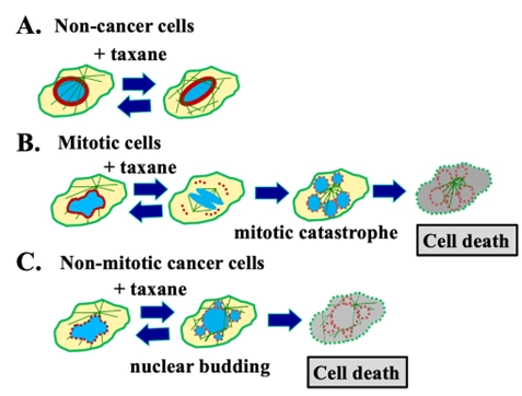
The paramount prerequisite for effective anti-cancer drugs is their ability to eradicate malignant cells while sparing non-cancer cells. The divergence in properties between malignant and non-cancer cells establishes a "therapeutic window," a critical consideration for achieving desirable treatment outcomes. Central to this is the imperative of a cancer drug's "selectivity and specificity." Taxanes, a pivotal class of successful anti-cancer drugs, continue to serve as the linchpin of cancer treatment due to their efficacy across a spectrum of cancer types. Operating as broad-spectrum chemotherapeutic agents, taxanes exert cytotoxic effects on proliferative cancer cells by binding to and stabilizing microtubules, disrupting mitosis, inducing mitotic catastrophe, and resulting in cell death. The distinct proliferative nature of cancer cells, as opposed to less proliferative non-cancer cells, affords taxanes a measure of specificity and selectivity. Nevertheless, sporadic yet recurring evidence suggests that taxanes also operate through non-mitotic mechanisms. Taxanes' binding and stabilization of microtubules lead to micronucleation and subsequent cell death, impacting both mitotic and non-mitotic cells. Recent discoveries indicate that the flexible and weakened nuclear envelope of malignant cells renders them sensitive to taxane-mediated micronucleation and cell death during various phases of the cell cycle. Conversely, non-cancerous cells typically exhibit a more robust and sturdy nuclear envelope, rendering them more tolerant to taxane-induced nuclear envelope fragmentation and subsequent micronucleation and cell death. The expression levels of nuclear envelope structural proteins, particularly Lamin A/C, emerge as indicators of taxane sensitivity. This evolving understanding underscores that nuclear envelope malleability, in conjunction with a high proliferation rate, is a pivotal determinant of taxane specificity and selectivity against malignant cells. These insights necessitate reconsidering oncological strategies to augment taxane efficacy, overcome resistance, and mitigate side effects.
Smith, E. R., Li, Z., Chen, Z., & Xu, X. (2024). Reassessing specificity/selectivity of taxane-based chemotherapy. Cancer Insight, 3(1), 27. doi:10.58567/ci03010002
ACS Style
Smith, E. R.; Li, Z.; Chen, Z.; Xu, X. Reassessing specificity/selectivity of taxane-based chemotherapy. Cancer Insight, 2024, 3, 27. doi:10.58567/ci03010002
AMA Style
Smith E R, Li Z, Chen Z et al.. Reassessing specificity/selectivity of taxane-based chemotherapy. Cancer Insight; 2024, 3(1):27. doi:10.58567/ci03010002
Chicago/Turabian Style
Smith, Elizabeth R.; Li, Zheshen; Chen, Zhe-Sheng; Xu, Xiang-Xi 2024. "Reassessing specificity/selectivity of taxane-based chemotherapy" Cancer Insight 3, no.1:27. doi:10.58567/ci03010002
NIH-NCI (R01 CA79716)
,
CDMRP DoD Concept and Pilot Awards (LC220190)
,
CDMRP DoD Concept and Pilot Awards (OC170318)
,
CDMRP DoD Concept and Pilot Awards (BC097189)
,
CDMRP DoD Concept and Pilot Awards (BC076832)
Share and Cite
ACS Style
Smith, E. R.; Li, Z.; Chen, Z.; Xu, X. Reassessing specificity/selectivity of taxane-based chemotherapy. Cancer Insight, 2024, 3, 27. doi:10.58567/ci03010002
AMA Style
Smith E R, Li Z, Chen Z et al.. Reassessing specificity/selectivity of taxane-based chemotherapy. Cancer Insight; 2024, 3(1):27. doi:10.58567/ci03010002
Chicago/Turabian Style
Smith, Elizabeth R.; Li, Zheshen; Chen, Zhe-Sheng; Xu, Xiang-Xi 2024. "Reassessing specificity/selectivity of taxane-based chemotherapy" Cancer Insight 3, no.1:27. doi:10.58567/ci03010002
APA style
Smith, E. R., Li, Z., Chen, Z., & Xu, X. (2024). Reassessing specificity/selectivity of taxane-based chemotherapy. Cancer Insight, 3(1), 27. doi:10.58567/ci03010002
Article Metrics
Article Access Statistics
References
Runowicz CD, Wiernik PH, Einzig AI, Goldberg GL, Horwitz SB: Taxol in ovarian cancer. Cancer 1993, 71(4 Suppl):1591-1596. https://doi.org/10.1002/cncr.2820710442
Rowinsky EK, Donehower RC: Paclitaxel (taxol). N Engl J Med 1995, 332(15):1004-1014. https://doi.org/10.1056/NEJM199504133321507
Holmes FA: Paclitaxel combination therapy in the treatment of metastatic breast cancer: a review. Semin Oncol 1996, 23(5 Suppl 11):46-56.
Friedrich M, Diesing D, Villena-Heinsen C, Felberbaum R, Kolberg HC, Diedrich K: Taxanes in the first-line chemotherapy of metastatic breast cancer: review. Eur J Gynaecol Oncol 2004, 25(1):66-70.
Hanna N: Current Standards and Clinical Trials in Systemic Therapy for Stage III Lung Cancer: What Is New? Am Soc Clin Oncol Educ Book 2015:e442-447. https://doi.org/10.14694/EdBook_AM.2015.35.e442
Bookman MA: Optimal primary therapy of ovarian cancer. Ann Oncol 2016, 27 Suppl 1:i58-i62. https://doi.org/10.1093/annonc/mdw088
Mosca L, Ilari A, Fazi F, Assaraf YG, Colotti G: Taxanes in cancer treatment: Activity, chemoresistance and its overcoming. Drug Resist Updat 2021, 54:100742. https://doi.org/10.1016/j.drup.2020.100742
Thomas H, Rosenberg P: Role of weekly paclitaxel in the treatment of advanced ovarian cancer. Crit Rev Oncol Hematol 2002, 44 Suppl:S43-51. https://doi.org/10.1016/s1040-8428(02)00103-8
Baird RD, Tan DS, Kaye SB: Weekly paclitaxel in the treatment of recurrent ovarian cancer. Nat Rev Clin Oncol 2010, 7(10):575-582. https://doi.org/10.1038/nrclinonc.2010.120
Baker VV: Salvage therapy for recurrent epithelial ovarian cancer. Hematol Oncol Clin North Am 2003, 17(4):977-988. https://doi.org/10.1016/s0889-8588(03)00057-1
Desai A, Mitchison TJ. Microtubule polymerization dynamics. Annu Rev Cell Dev Biol. 1997; 13:83-117.https://doi.org/10.1146/annurev.cellbio.13.1.83
Blagosklonny MV, Fojo T. Molecular effects of paclitaxel: myths and reality (a critical review). Int J Cancer 1999 Oct 8; 83(2):151-6. https://doi.org/10.1002/(SICI)1097-0215(19991008)83:2%3C151::AID-IJC1%3E3.0.CO;2-5
Visconti R, Grieco D. Fighting tubulin-targeting anticancer drug toxicity and resistance. Endocr Relat Cancer 2017 Sep; 24(9):T107-T17. https://doi.org/10.1530/ERC-17-0120
Barbuti AM, Chen ZS. Paclitaxel through the ages of anticancer therapy: Exploring its role in chemoresistance and radiation therapy. Cancers (Basel) 2015; 7(4):2360-71. https://doi.org/10.3390/cancers7040897
Gallego-Jara J, Lozano-Terol G, Sola-Martínez RA, Cánovas-Díaz M, de Diego Puente, T. A compressive review about taxol: History and future challenges. Molecules 2020; 25(24): 5986.
https://doi.org/10.3390/molecules25245986
Schiff PB, Fant J, Horwitz SB. Promotion of microtubule assembly in vitro by taxol. Nature 1979 Feb 22;277(5698):665-7. https://doi.org/10.1038/277665a0
Schiff PB, Horwitz SB. Taxol stabilizes microtubules in mouse fibroblast cells. Proc Natl Acad Sci U S A. 1980 Mar;77(3):1561-5. https://doi.org/10.1073/pnas.77.3.1561
Jordan MA. Mechanism of action of antitumor drugs that interact with microtubules and tubulin. Curr Med Chem Anticancer Agents 2002;2(1):1-17. https://doi.org/10.2174/1568011023354290
Jordan MA, Wilson L. Microtubules as a target for anticancer drugs. Nat Rev Cancer 2004; 4:253-65. https://doi.org/10.1038/nrc1317
Smith ER, Leal J, Amaya C, Li B, Xu XX. Nuclear Lamin A/C Expression Is a Key Determinant of Paclitaxel Sensitivity. Mol Cell Biol. 2021 Jun 23;41(7):e0064820. https://doi.org/10.1128/MCB.00648-20
Smith ER, Wang JQ, Yang DH, Xu XX. Paclitaxel resistance related to nuclear envelope structural sturdiness. Drug Resist Updat. 2022 Dec;65:100881. https://doi.org/10.1016/j.drup.2022.100881
Yang CH, Horwitz SB. Taxol®: The First Microtubule Stabilizing Agent. Int J Mol Sci. 2017 Aug 9;18(8). https://doi.org/10.3390/ijms18081733
Manfredi JJ, Parness J, Horwitz SB. Taxol binds to cellular microtubules. J Cell Biol. 1982; 94(3):688-96. https://doi.org/10.1083/jcb.94.3.688
Diaz JF, Andreu JM. Assembly of purified GDP-tubulin into microtubules induced by taxol and taxotere: reversibility, ligand stoichiometry, and competition. Biochemistry 1993; 32:2747-2755. https://doi.org/10.1021/bi00062a003
Michalakis J, Georgatos SD, de Bree E, Polioudaki H, Romanos J, Georgoulias V, Tsiftsis DD, Theodoropoulos PA. Short-term exposure of cancer cells to micromolar doses of paclitaxel, with or without hyperthermia, induces long-term inhibition of cell proliferation and cell death in vitro. Ann Surg Oncol. 2007; 14(3):1220-1228. https://doi.org/10.1245/s10434-006-9305-4
Koshiba H, Hosokawa K, Mori T, Kubo A, Watanabe A, Honjo H. Intravenous paclitaxel is specifically retained in human gynecologic carcinoma tissues in vivo. Int J Gynecol Cancer 2009; 19(4): 484-4488. https://doi.org/10.1111/IGC.0b013e3181a130db
Mori T, Kinoshita Y, Watanabe A, Yamaguchi T, Hosokawa K, Honjo H. Retention of paclitaxel in cancer cells for 1 week in vivo and in vitro. Cancer Chemother Pharmacol. 2006; 58(5): 665-672. https://doi.org/10.1007/s00280-006-0209-6
Wiernik PH, Schwartz EL, Strauman JJ, Dutcher JP, Lipton RB, Paietta E. Phase I clinical and pharmacokinetic study of taxol. Cancer Res. 1987; 47(9):2486-93.
Smith ER, Xu XX. Breaking malignant nuclei as a non-mitotic mechanism of taxol/paclitaxel. J Cancer Biol. 2021;2(4):86-93. https://doi.org/10.46439/cancerbiology.2.031
Mitchison TJ. Microtubule dynamics and kinetochore function in mitosis. Annu Rev Cell Biol. 1988;4:527-49. https://doi.org/10.1146/annurev.cb.04.110188.002523
Kline-Smith SL, Walczak CE. Mitotic spindle assembly and chromosome segregation: refocusing on microtubule dynamics. Mol Cell. 2004 Aug 13;15(3):317-27.https://doi.org/10.1016/j.molcel.2004.07.012
Jordan MA, Toso RJ, Thrower D, Wilson L. Mechanism of mitotic block and inhibition of cell proliferation by taxol at low concentrations. Proc Natl Acad Sci. USA 1993; 90:9552-9556. https://doi.org/10.1073/pnas.90.20.9552
Horwitz SB. Taxol (paclitaxel): mechanisms of action. Ann Oncol. 1994;5 Suppl 6:S3-6.
Shi J, Mitchison TJ. Cell death response to anti-mitotic drug treatment in cell culture, mouse tumor model and the clinic. Endocr Relat Cancer 2017 Sep;24(9):T83-T96. https://doi.org/10.1530/ERC-17-0003
Morse DL, Gray H, Payne CM, Gillies RJ. Docetaxel induces cell death through mitotic catastrophe in human breast cancer cells. Mol Cancer Ther. 2005; 4(10):1495-504. https://doi.org/10.1158/1535-7163.MCT-05-0130
Weaver BA. How Taxol/paclitaxel kills cancer cells. Mol Biol Cell 2014;25(18):2677-81. https://doi.org/10.1091/mbc.E14-04-0916
Zasadil LM, Andersen KA, Yeum D, Rocque GB, Wilke LG, Tevaarwerk AJ, Raines RT, Burkard ME, Weaver BA. Cytotoxicity of paclitaxel in breast cancer is due to chromosome missegregation on multipolar spindles. Sci Transl Med. 2014; 6(229):229ra43.https://doi.org/10.1126/scitranslmed.3007965
Zhu Y, Zhou Y, Shi J. Post-slippage multinucleation renders cytotoxic variation in anti-mitotic drugs that target the microtubules or mitotic spindle. Cell Cycle 2014; 13(11): 1756-64.https://doi.org/10.4161/cc.28672
Giannakakou P, Nakano M, Nicolaou KC, O'Brate A, Yu J, Blagosklonny MV, Greber UF, Fojo T. Enhanced microtubule-dependent trafficking and p53 nuclear accumulation by suppression of microtubule dynamics. Proc Natl Acad Sci USA. 2002; 99(16):10855-10860.https://doi.org/10.1073/pnas.132275599
Bouchet BP, Akhmanova A. Microtubules in 3D cell motility. J Cell Sci. 2017 Jan 1; 130(1):39-50. https://doi.org/10.1242/jcs.189431
Gascoigne K, Taylor SS. How do anti-mitotic drugs kill cancer cells. J Cell Sci. 2009; 122:2579-85. https://doi.org/10.1242/jcs.039719
Shi J, Orth JD, Mitchison T. Cell type variation in responses to antimitotic drugs that target microtubules and kinesin-5. Cancer Res. 2008 May 1;68(9):3269-76. https://doi.org/10.1158/0008-5472.CAN-07-6699
Mielke S, Sparreboom A, Mross K. Peripheral neuropathy: a persisting challenge in paclitaxel-based regimes. Eur. J. Cancer 2006; 42(1):24-30. https://doi.org/10.1016/j.ejca.2005.06.030
Franker MA, Hoogenraad CC. Microtubule-based transport - basic mechanisms, traffic rules and role in neurological pathogenesis. J Cell Sci. 2013 Jun 1;126(Pt 11):2319-29. https://doi.org/10.1242/jcs.115030
Gornstein E, Schwarz TL. The paradox of paclitaxel neurotoxicity: Mechanisms and unanswered questions. Neuropharmacology 2014;76 Pt A:175-83. https://doi.org/10.1016/j.neuropharm.2013.08.016
Komlodi-Pasztor E, Sackett DL, Fojo AT. Inhibitors targeting mitosis: tales of how great drugs against a promising target were brought down by a flawed rationale. Clin Cancer Res. 2012; 18(1):51-63. https://doi.org/10.1038/nrclinonc.2010.228
Schimming R, Mason KA, Hunter N, Weil M, Kishi K, Milas L. Lack of correlation between mitotic arrest or apoptosis and antitumor effect of docetaxel. Cancer Chemother Pharmacol. 1999;43(2):165-72. https://doi.org/10.1007/s002800050879
Yan VC, Butterfield HE, Poral AH, Yan MJ, Yang KL, Pham CD, Muller FL. Why great mitotic inhibitors make poor cancer drugs. Trends Cancer 2020; 6: 924-941. https://doi.org/10.1016/j.trecan.2020.05.010
Mitchison TJ. The proliferation rate paradox in antimitotic chemotherapy. Mol Biol Cell 2012 Jan; 23(1):1-6. https://doi.org/10.1091/mbc.E10-04-0335
Komlodi-Pasztor E, Sackett D, Wilkerson J, Fojo T. Mitosis is not a key target of microtubule agents in patient tumors. Nat Rev Clin Oncol. 2011; 8(4):244-50. https://doi.org/10.1038/nrclinonc.2010.228
Fürst R, Vollmar AM. A new perspective on old drugs: non-mitotic actions of tubulin-binding drugs play a major role in cancer treatment. Pharmazie. 2013 Jul;68(7):478-83.
Field JJ, Kanakkanthara A, Miller JH:Microtubule-targeting agents are clinically successful due to both mitotic and interphase impairment of microtubule function. Bioorg Med Chem. 2014; 22(18):5050-9. https://doi.org/10.1016/j.bmc.2014.02.035
Merlin JL, Bour-Dill C, Marchal S, Bastien L, Gramain MP. Resistance to paclitaxel induces time-delayed multinucleation and DNA fragmentation into large fragments in MCF-7 human breast adenocarcinoma cells. Anti-Cancer Drugs 2000; 11:295-302. https://doi.org/10.1097/00001813-200004000-00011
Mitchison TJ, Pineda J, Shi J, Florian S. Is inflammatory micronucleation the key to a successful anti-mitotic cancer drug? Open Biol. 2017 Nov; 7(11):170182. https://doi.org/10.1098/rsob.170182
Panvichian R, Orth K, Day ML, Day KC, Pilat MJ, Pienta KJ. Paclitaxel-associated multimininucleation is permitted by the inhibition of caspase activation: a potential early step in drug resistance. Cancer Res. 1998; 58:4667-72.
Gjyrezi A, Xie F, Voznesensky O, Khanna P, Calagua C, Bai Y, Kung J, Wu J, Corey E, Montgomery B, Mace S, Gianolio DA, Bubley GJ, Balk SP, Giannakakou P, Bhatt RS. Taxane resistance in prostate cancer is mediated by decreased drug-target engagement. J Clin Invest. 2020 Jun 1;130(6):3287-3298. https://doi.org/10.1172/JCI132184
Vargas JD, Hatch EM, Anderson DJ, Hetzer MW. Transient nuclear envelope rupturing during interphase in human cancer cells. Nucleus 2012; 3(1):88-100. https://doi.org/10.4161/nucl.18954
Fojo T, Menefee M. Mechanisms of multidrug resistance: the potential role of microtubule-stabilizing agents. Ann Oncol. 2007 Jul;18 Suppl 5:v3-8. https://doi.org/10.1093/annonc/mdm172
Moss SF, Krivosheyev V, de Souza A, Chin K, Gaetz HP, Chaudhary N, Worman HJ, Holt PR. Decreased and aberrant nuclear lamin expression in gastrointestinal tract neoplasms. Gut 1999; 45:723-9. https://doi.org/10.1136/gut.45.5.723
Wu Z, Wu L, Weng D, Xu D, Geng J, Zhao F. Reduced expression of lamin A/C correlates with poor histological differentiation and prognosis in primary gastric carcinoma. J Exp Clin Cancer Res. 2009; 28:8. https://doi.org/10.1186/1756-9966-28-8
Capo-chichi CD, Cai KQ, Smedberg J, Ganjei-Azar P, Godwin AK, Xu XX. Loss of A-type lamin expression compromises nuclear envelope integrity in breast cancer. Chin J Cancer 2011a;30:415-25. https://doi.org/10.5732/cjc.010.10566
Capo-chichi CD, Cai KQ, Simpkins F, Ganjei-Azar P, Godwin AK, Xu XX. Nuclear envelope structural defects cause chromosomal numerical instability and aneuploidy in ovarian cancer. BMC Med. 2011b; 9:28. https://doi.org/10.1186/1741-7015-9-28
Foster CR, Przyborski SA, Wilson RG, Hutchison CJ. Lamins as cancer biomarkers. Biochem Soc Trans. 2010;38:297-300. https://doi.org/10.1042/BST0380297
Willis ND, Cox TR, Rahman-Casañs SF, Smits K, Przyborski SA, van den Brandt P, van Engeland M, Weijenberg M, Wilson RG, de Bruïne A, Hutchison CJ. Lamin A/C is a risk biomarker in colorectal cancer. PLoS One 2008;3:e2988. https://doi.org/10.1371/journal.pone.0002988
Smith ER, George SH, Kobetz E, Xu XX. New biological research and understanding of Papanicolaou's test. Diagn Cytopathol. 2018 Jun;46(6):507-15. https://doi.org/10.1002/dc.23941
Smith ER, Meng Y, Moore R, Tse JD, Xu AG, Xu XX. Nuclear envelope structural proteins facilitate nuclear shape changes accompanying embryonic differentiation and fidelity of gene expression. BMC Cell Biol. 2017 Jan 14;18(1):8. https://doi.org/10.1186/s12860-017-0125-0
Zink D, Fischer AH, Nickerson JA. Nuclear structure in cancer cells. Nat Rev Cancer. 2004 Sep;4(9):677-87. https://doi.org/10.1038/nrc1430
Capo-Chichi CD, Yeasky TM, Smith ER, Xu XX. Nuclear envelope structural defect underlies the main cause of aneuploidy in ovarian carcinogenesis. BMC Cell Biol. 2016 Nov 22;17(1):37. https://doi.org/10.1186/s12860-016-0114-8
Shah P, Wolf K, Lammerding J. Bursting the Bubble - Nuclear Envelope Rupture as a Path to Genomic Instability? Trends Cell Biol. 2017 Aug;27(8):546-55. https://doi.org/10.1016/j.tcb.2017.02.008
Maciejowski J, Hatch EM. Nuclear Membrane Rupture and Its Consequences. Annu Rev Cell Dev Biol. 2020 Oct 6;36:85-114. https://doi.org/10.1146/annurev-cellbio-020520-120627